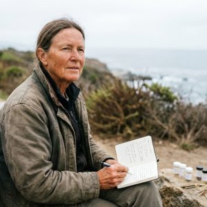
EXERCISE BRAIN AGING research examines how physical activity relates to neuroprotection, synaptic plasticity, and resilience against age-related cognitive decline.
This overview maps mechanisms, summarizes human and experimental evidence, and highlights uncertainties to support careful interpretation without prescriptive claims.
Mechanistic Pathways Linking Exercise to Neuroprotection
Multiple, interacting biological systems are implicated in how exercise may influence the aging brain:
Neurotrophin signaling and synaptic plasticity: Physical activity is associated with increased brain-derived neurotrophic factor (BDNF) signaling through TrkB and downstream CREB activity, which supports synaptic plasticity and hippocampal neurogenesis in experimental models.
Muscle-derived myokines, including irisin (cleaved from FNDC5) and cathepsin B, have been proposed as peripheral signals that may modulate neuronal BDNF expression; these mechanisms remain under investigation in humans.
Mitochondrial health and cellular stress responses: Exercise engages mitochondrial biogenesis via PGC‑1α, NRF1/2, and TFAM, and may influence mitophagy pathways (e.g., PINK1/Parkin), ROS handling, and mitochondrial dynamics (MFN1/2, OPA1, DRP1) in model systems.
Readers can explore related mechanisms in exercise-driven mitochondrial adaptations in aging, AMPK longevity pathway, and mTOR signaling in aging.
Metabolic and nutrient-sensing pathways: Improved systemic insulin sensitivity and altered lactate signaling (astrocyte–neuron shuttling through MCTs) may influence neuronal metabolism.
These processes intersect with insulin signaling and aging and nutrient-sensing aging networks.
Vascular and angiogenic effects: Exercise can increase cerebral perfusion and is associated with angiogenic signaling (e.g., IGF‑1, VEGF) in experimental models, which may support neurogenesis niches.
Maintenance of blood–brain barrier integrity and perivascular function is under active study.
Neuroimmune modulation: Inflammation may be attenuated through shifts in microglial activation states and cytokine profiles in preclinical models, potentially reducing chronic neuroinflammation that accompanies aging.
For broader context, see inflammation and aging link and cellular senescence in neurodegeneration.
Circuit-level organization: Functional changes observed in humans include task-related activity in hippocampal and prefrontal circuits and connectivity within large-scale networks; results vary by study design, imaging modality, and participant characteristics.
Human Observational Evidence
Epidemiologic cohorts generally report that higher habitual physical activity is associated with reduced risk of cognitive decline and incident dementia.
These associations are subject to confounding (e.g., socioeconomic status, baseline health) and potential reverse causation (subclinical neurodegeneration may reduce activity years before diagnosis).
Exposure measurement (self-report versus devices), activity domains (leisure, occupational, transport), and cohort age structure contribute to heterogeneity.
Human Interventional Trials
Cognition and brain structure: Randomized and controlled exercise trials in older adults have reported modest improvements in executive function and memory.
Some studies indicate hippocampal volume increases on MRI after structured aerobic training over several months. Effects are not universal and vary by duration, intensity, adherence, and cognitive status at baseline.
Biomarkers: Acute exercise often increases circulating BDNF transiently; whether peripheral BDNF reflects central concentrations remains uncertain.
Imaging endpoints (e.g., regional volumes, perfusion, and connectivity) and fluid biomarkers (inflammatory cytokines, neurotrophins) are being used to characterize brain responses to training.
Heterogeneity: Trials differ in modality (aerobic, resistance, mixed), intensity, session structure, and supervision, limiting cross-study comparability.
Studies in mild cognitive impairment and early neurodegenerative disease are ongoing and report mixed outcomes.
Animal and Cellular Models
In rodents, voluntary wheel running and treadmill paradigms increase hippocampal neurogenesis, dendritic complexity, synaptic plasticity markers, and neurotrophin levels.
Disease-model studies often suggest delayed pathology or improved behavior with regular activity; translation to human disease progression remains an open question due to species differences, controlled environments, and effect sizes that may not generalize.
Intensity, Modality, and Recovery Considerations
Evidence on optimal intensity and modality for neuroprotection is mixed. Aerobic and resistance training can each engage neurotrophin and vascular pathways to varying degrees.
Excessive training loads may increase stress hormones and inflammatory signals; see exercise intensity and longevity pathways and overtraining and aging risk in the nervous system for context on potential trade-offs.
Interactions with Sleep, Circadian Timing, and Environment
Sleep and circadian systems shape neuroplasticity, immune responses, and metabolic regulation relevant to brain aging.
Exercise timing relative to circadian rhythm and sleep quality is under investigation; for related coverage, see sleep patterns and longevity and circadian rhythm and aging.
Environmental exposures and psychosocial stress may also modify exercise–brain relationships; see psychological stress and aging and stress recovery and aging.
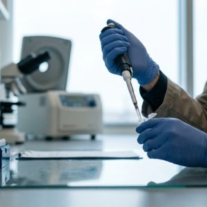
Measurement, Biomarkers, and Aging Clocks
Imaging: MRI-based hippocampal and cortical volumes, diffusion metrics, cerebral blood flow, and network connectivity are frequently used to track neurobiological change.
Circulating markers: Peripheral BDNF, IGF‑1, VEGF, inflammatory cytokines, and myokines are studied as proxies; their relationship to central processes is not one-to-one.
Multi-omic and epigenetic readouts: DNA methylation signatures and transcriptional changes are under study to quantify biological aging in the brain and periphery.
See biological aging markers for the brain, measuring biological age, epigenetic aging markers in neural tissue, DNA methylation aging clocks, and limits of epigenetic reversal in the brain.
Disease-Specific Context and Emerging Directions
For Alzheimer’s and Parkinson’s disease, exercise is being studied as a potential adjunct to standard care, with endpoints that include cognitive tests, motor scales, imaging biomarkers, and fluid markers (amyloid, tau, α‑synuclein) where applicable.
Current evidence does not establish prevention or disease modification, although some trials report functional or biomarker signals.
Related reporting can be found in brain tissue regeneration research updates and Alzheimer’s brain stimulation news.
Equity, Culture, and Policy Interfaces
Access to safe environments for movement, green space, and community resources may shape the capacity to engage in activity linked with neuroprotection.
Policy discussions on infrastructure and aging intersect with this topic; see global longevity policy for equitable brain health.
Cultural narratives can distort expectations about rapid brain changes; for a systems view of interacting factors, see systems biology of aging networks and biological resilience in aging.
Bibliographic References
- Erickson, Kirk I., Michelle W. Voss, Ruchika S. Prakash, et al. “Exercise Training Increases Size of Hippocampus and Improves Memory.” Proceedings of the National Academy of Sciences 108, no. 7 (2011): 3017-3022. https://doi.org/10.1073/pnas.1015950108
- Hillman, Charles H., Kirk I. Erickson, and Arthur F. Kramer. “Be Smart, Exercise Your Heart: Exercise Effects on Brain and Cognition.” Nature Reviews Neuroscience 9, no. 1 (2008): 58-65. https://doi.org/10.1038/nrn2298
- Cotman, Carl W., and Nicole C. Berchtold. “Exercise: A Behavioral Intervention to Enhance Brain Health and Plasticity.” Trends in Neurosciences 25, no. 6 (2002): 295-301. https://doi.org/10.1016/S0166-2236(02)02143-4
Why this Matters to People
Imagine your brain is like a garden. When you move and play, it’s like watering and feeding the plants.
Exercise helps keep the «plants» (your brain cells) strong and growing, so they can do their jobs better, like remembering things and solving puzzles. This means you can pay attention in school and remember what your friends say.
If you make moving a habit – like biking, hopping, swimming, or even walking the dog – these actions could keep your brain sharper as you grow up. It also means you might avoid forgetting things when you’re older.
Your daily fun movement, even simple games, makes your brain healthier, so you feel happier and have more energy for learning and all your favorite activities!
FAQs
How Might Exercise Influence Neuroprotection in Aging?
Studies suggest that physical activity engages neurotrophin signaling (BDNF), vascular support, mitochondrial biogenesis, and neuroimmune modulation. These effects are most consistently demonstrated in animal and cellular models, with variable findings in human trials.
Which Brain Regions Are Most Often Reported to Change with Exercise?
Human imaging studies frequently examine the hippocampus and prefrontal cortex. Some trials report increases in hippocampal volume and improvements in executive function after structured training, while other studies find minimal or no change.
Is Higher-Intensity Exercise Superior for Brain Health?
Evidence is mixed and depends on modality, duration, and participant characteristics. Both moderate and higher intensities have been associated with neurobiological signals in different studies. For context, see exercise intensity and longevity pathways.
Does Exercise Prevent Alzheimer’s Disease?
Current evidence does not establish prevention. Observational research associates higher activity with lower risk, and trials are investigating cognitive and biomarker outcomes. Findings vary, and causality is not confirmed.
What Biomarkers Track Brain Responses to Exercise?
Common endpoints include MRI-based regional volumes, cerebral blood flow, task or resting-state connectivity, peripheral BDNF and IGF‑1, and inflammatory markers. Relationships between peripheral biomarkers and central brain biology are not direct and remain under active study.